Skip to content
Anatly

Sacrum: anatomy, landmarks and articulations

Quick answer

The sacrum is a triangular bone formed by five fused sacral vertebrae. It forms the posterior wall of the pelvis, articulates with L5 above, the coccyx below and the hip bones at the sacroiliac joints. Its foramina carry the sacral spinal nerves.

Key facts

TypeIrregular bone (fused vertebrae).
LocationPosterior pelvis, between the hip bones.
LandmarksBase, promontory, ala, anterior and posterior sacral foramina, median and lateral sacral crests, sacral hiatus, cornua.
ArticulationsFifth lumbar vertebra, coccyx and hip bones (sacroiliac joints).
Muscle attachmentsGluteus maximus, piriformis, erector spinae, multifidus, iliacus.
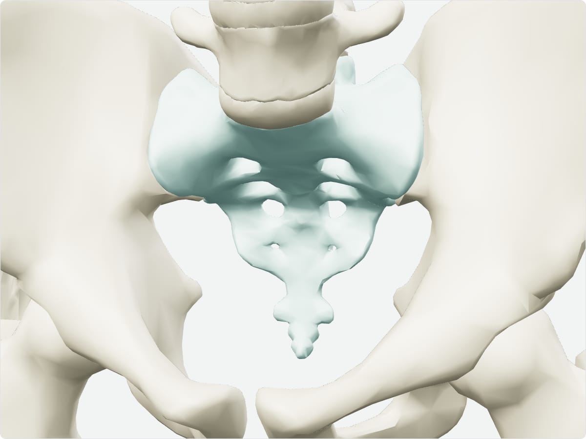
3D model of the sacrum shown with nearby bones

3D model: BodyParts3D (CC BY 4.0), adult male reference anatomy. Highlighted structure shown with nearby bones for context.

About the sacrum

The sacral promontory is used as a landmark in obstetrics. The sacral canal is the continuation of the vertebral canal and contains the cauda equina.

Clinical relevance

Frequently asked questions

How many vertebrae fuse to form the sacrum?

Five.

What passes through the sacral foramina?

The anterior and posterior rami of the sacral spinal nerves.

Test yourself

Back bone quiz

Practise origins, insertions, nerve supply and locations with instant feedback.

Explore in context

Open the sacrum in the full 3D atlas

Toggle body systems, explode the model and inspect every neighbouring structure.