Skip to content
Anatly

Hip bone: anatomy, landmarks and articulations

Also called: pelvic bone, os coxae, innominate bone

Quick answer

The hip bone, or os coxae, is a large irregular bone formed by the fusion of the ilium, ischium and pubis. The two hip bones meet at the pubic symphysis and articulate with the sacrum to form the pelvis. The acetabulum on its lateral surface receives the head of the femur.

Key facts

TypeIrregular bone formed by three fused bones.
LocationLateral wall of the pelvis.
LandmarksIliac crest, anterior and posterior superior iliac spines, acetabulum, obturator foramen, ischial tuberosity, ischial spine, pubic symphysis surface, greater and lesser sciatic notches.
ArticulationsSacrum (sacroiliac joint), opposite hip bone (pubic symphysis), femur (hip joint).
Muscle attachmentsGluteal muscles, hamstrings, adductors, sartorius, rectus femoris, iliacus and abdominal wall muscles.
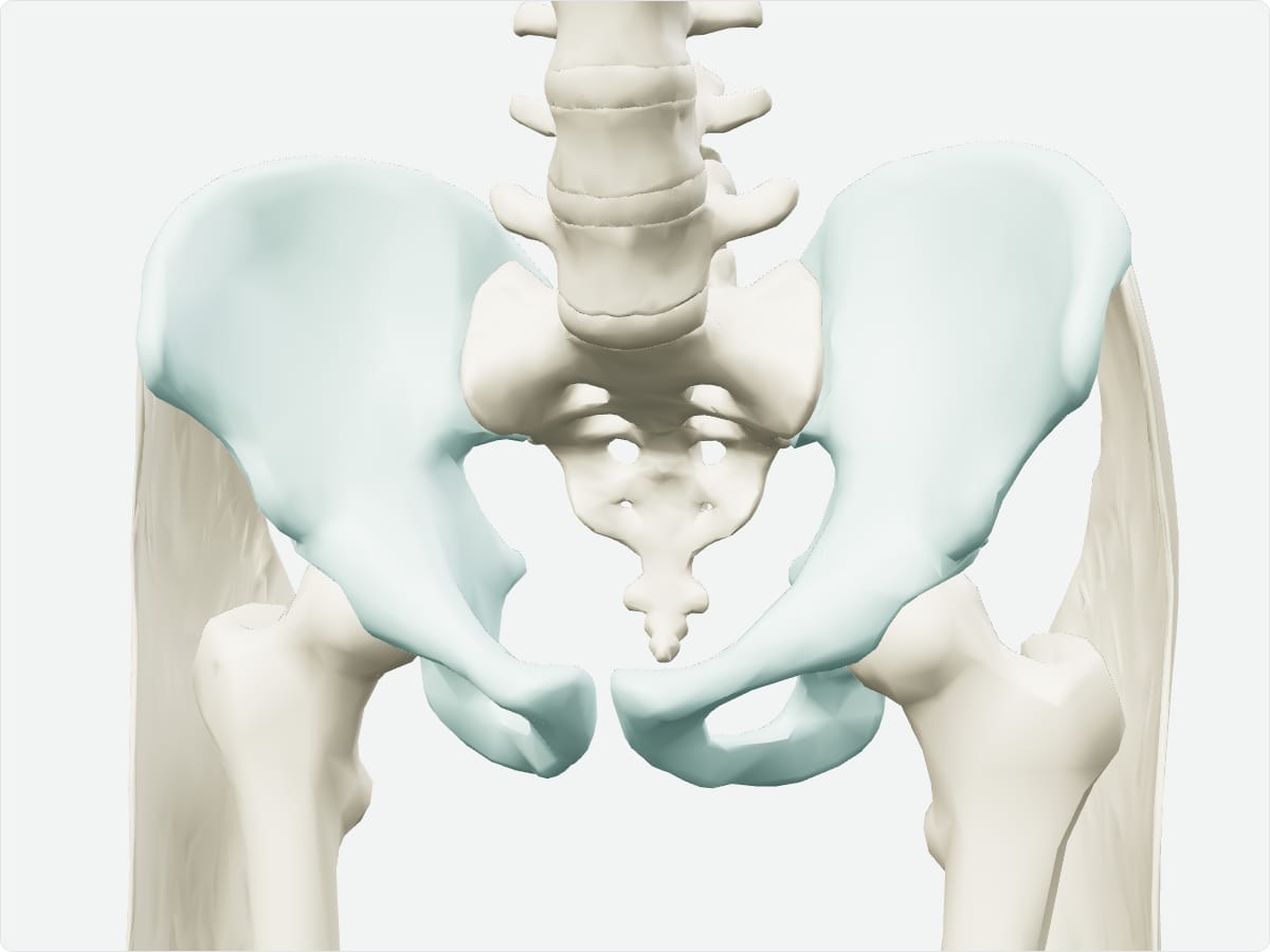
3D model of the hip bone shown with nearby bones

3D model: BodyParts3D (CC BY 4.0), adult male reference anatomy. Highlighted structure shown with nearby bones for context.

About the hip bone

The three bones fuse in the acetabulum during adolescence. The shape of the pelvis differs between sexes, related to childbirth.

Clinical relevance

Frequently asked questions

Which three bones form the hip bone?

The ilium, ischium and pubis.

What is the acetabulum?

The cup-shaped socket on the hip bone that receives the femoral head.

Test yourself

Abdomen and pelvis bone quiz

Practise origins, insertions, nerve supply and locations with instant feedback.

Explore in context

Open the hip bone in the full 3D atlas

Toggle body systems, explode the model and inspect every neighbouring structure.