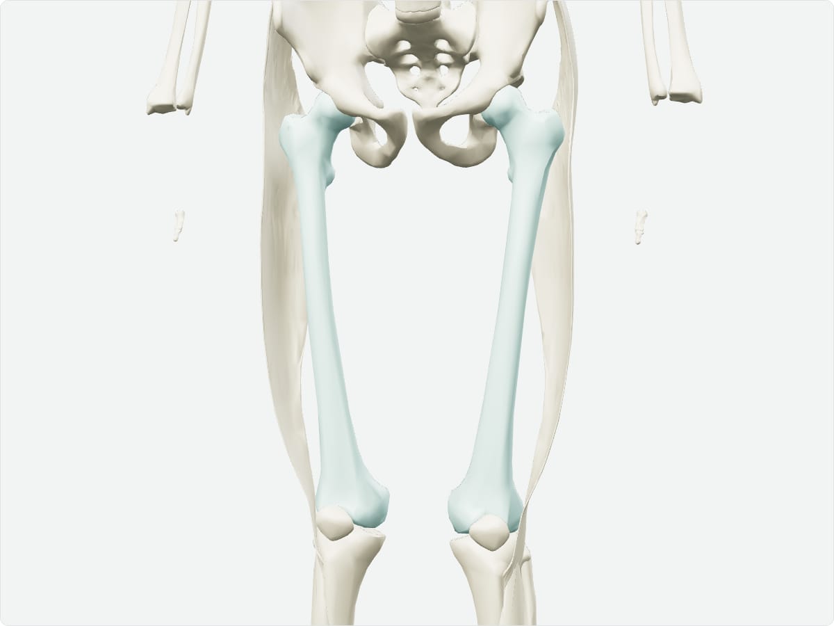
The femur is the thigh bone and the longest, strongest bone of the body. Its head articulates with the acetabulum of the hip bone, and its distal condyles form the knee joint with the tibia and patella. Major landmarks include the neck, greater and lesser trochanters, the linea aspera and the adductor tubercle.
Key facts
Type
Long bone.
Location
Thigh, between the hip and knee.
Landmarks
Head, fovea, neck, greater and lesser trochanters, intertrochanteric line and crest, linea aspera, gluteal tuberosity, medial and lateral condyles, epicondyles, adductor tubercle, patellar surface.
Articulations
Hip bone (hip joint); tibia and patella (knee joint).
Muscle attachments
Gluteal muscles, iliopsoas, adductors, quadriceps (vasti), short head of biceps femoris, gastrocnemius, popliteus.
3D model: BodyParts3D (CC BY 4.0), adult male reference anatomy. Highlighted structure shown with nearby bones for context.
About the femur
The neck of the femur meets the shaft at an angle of about 125 degrees in adults. This angle, together with the blood supply of the femoral head, explains why femoral neck fractures are serious.
Clinical relevance
Femoral neck fractures in older adults can disrupt the blood supply to the head and lead to avascular necrosis.
The femur is the site of many muscular avulsion injuries in adolescents.
Frequently asked questions
Why is a femoral neck fracture serious?
The femoral head depends on small vessels that run along the neck, so a fracture can cause avascular necrosis.