About the tensor fasciae latae
With the upper fibres of gluteus maximus it tensions the iliotibial tract, which helps keep the knee extended when standing.
Clinical relevance
- Iliotibial band syndrome causes lateral knee pain in runners and cyclists.
Also called: TFL
The tensor fasciae latae is a small muscle on the upper lateral thigh. It arises from the anterior superior iliac spine and inserts into the iliotibial tract, which attaches to the lateral tibial condyle. The superior gluteal nerve supplies it, and it flexes, abducts and medially rotates the hip while tensing the iliotibial band.
| Origin | Anterior superior iliac spine and the anterior part of the outer lip of the iliac crest. |
|---|---|
| Insertion | Iliotibial tract, which attaches to the lateral tibial condyle (Gerdy's tubercle). |
| Innervation | Superior gluteal nerve (L4-L5). |
| Blood supply | Ascending branch of the lateral circumflex femoral artery. |
| Action | Flexes, abducts and medially rotates the thigh; tenses the iliotibial tract to stabilise the knee and pelvis. |
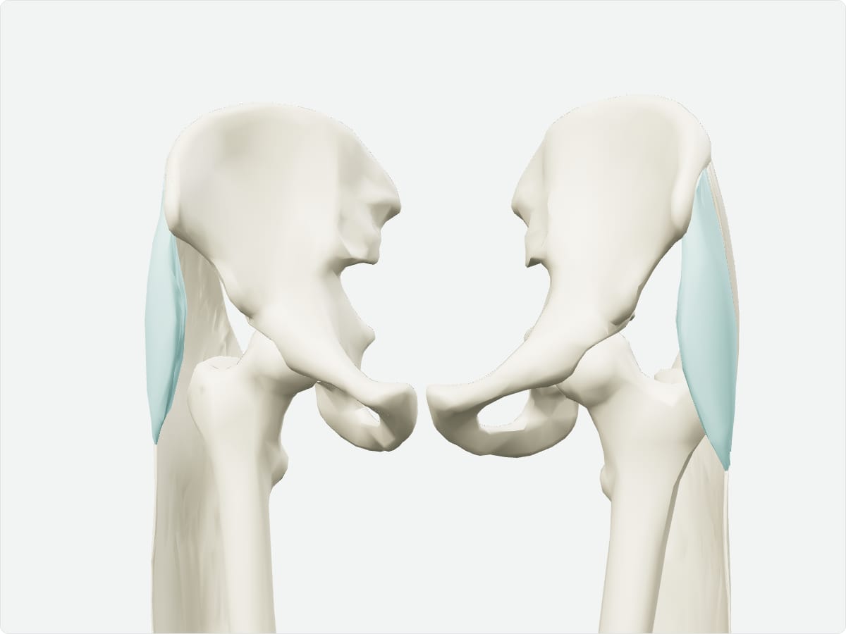
3D model: BodyParts3D (CC BY 4.0), adult male reference anatomy. Highlighted structure shown with nearby bones for context.
With the upper fibres of gluteus maximus it tensions the iliotibial tract, which helps keep the knee extended when standing.
It arises from the anterior superior iliac spine and attaches to the iliotibial tract.
The superior gluteal nerve.
Test yourself
Lower limb muscle quiz
Practise origins, insertions, nerve supply and locations with instant feedback.
Explore in context
Open the tensor fasciae latae in the full 3D atlas
Toggle body systems, explode the model and inspect every neighbouring structure.