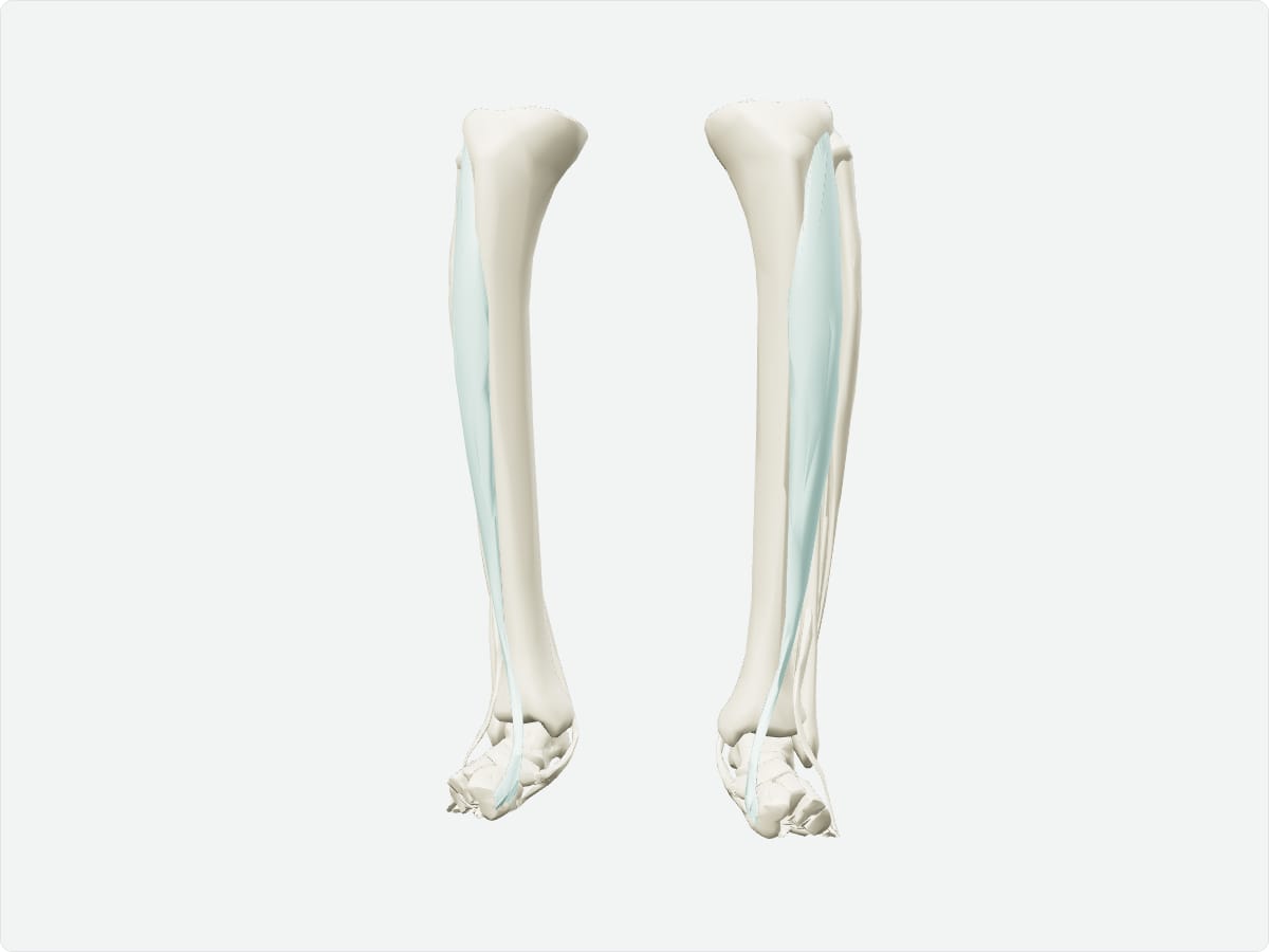
Tibialis anterior: origin, insertion, nerve supply and action
Quick answer
The tibialis anterior is the large muscle lateral to the shin bone in the anterior compartment of the leg. It arises from the lateral tibia and interosseous membrane and inserts on the medial cuneiform and base of the first metatarsal. The deep fibular nerve supplies it, and it dorsiflexes and inverts the foot.
Key facts
Origin
Lateral condyle and upper two thirds of the lateral surface of the tibia and the interosseous membrane.
Insertion
Medial and plantar surfaces of the medial cuneiform and base of the first metatarsal.
Innervation
Deep fibular (peroneal) nerve (L4-L5).
Blood supply
Anterior tibial artery.
Action
Dorsiflexes the ankle and inverts the foot; helps maintain the medial longitudinal arch.
3D model: BodyParts3D (CC BY 4.0), adult male reference anatomy. Highlighted structure shown with nearby bones for context.
About the tibialis anterior
It lifts the front of the foot during the swing phase of walking so that the toes clear the ground.
Clinical relevance
Weakness of tibialis anterior due to deep fibular nerve or L4-L5 injury causes foot drop.
Overuse of the muscle is a common cause of shin splints.
Frequently asked questions
What is foot drop?
Inability to dorsiflex the foot, usually due to weakness of tibialis anterior from deep or common fibular nerve injury or an L4-L5 lesion.
What movements does tibialis anterior produce?
Dorsiflexion of the ankle and inversion of the foot.