Skip to content
Anatly

Fibula: anatomy, landmarks and articulations

Also called: calf bone

Quick answer

The fibula is the slender lateral bone of the leg. It carries little body weight but provides attachment for muscles and forms the lateral malleolus, which stabilises the ankle joint. The common fibular nerve winds around its neck just below the knee.

Key facts

TypeLong bone.
LocationLateral leg, parallel to the tibia.
LandmarksHead, neck, shaft, lateral malleolus, malleolar fossa.
ArticulationsTibia (superior and inferior tibiofibular joints) and talus (ankle joint).
Muscle attachmentsBiceps femoris, fibularis muscles, soleus, tibialis posterior, extensor digitorum longus.
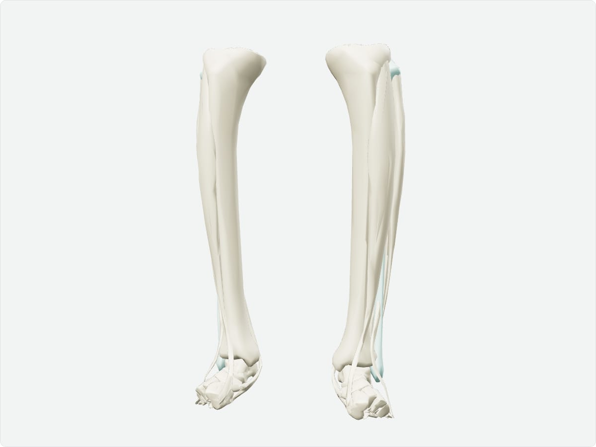
3D model of the fibula shown with nearby bones

3D model: BodyParts3D (CC BY 4.0), adult male reference anatomy. Highlighted structure shown with nearby bones for context.

About the fibula

The fibula stabilises the ankle by forming the lateral wall of the mortise that grips the talus.

Clinical relevance

Frequently asked questions

Does the fibula bear weight?

Only a small fraction; most weight is transmitted through the tibia.

Which nerve is at risk near the fibular neck?

The common fibular (peroneal) nerve.

Test yourself

Lower limb bone quiz

Practise origins, insertions, nerve supply and locations with instant feedback.

Explore in context

Open the fibula in the full 3D atlas

Toggle body systems, explode the model and inspect every neighbouring structure.