Popliteus: origin, insertion, nerve supply and action
Quick answer
The popliteus is a small triangular muscle at the back of the knee. It arises from the lateral femoral condyle and inserts on the posterior tibia above the soleal line. The tibial nerve supplies it, and it unlocks the fully extended knee by rotating the femur laterally on the tibia.
Key facts
Origin
Lateral surface of the lateral femoral condyle, by an intracapsular tendon.
Insertion
Posterior surface of the tibia above the soleal line.
Innervation
Tibial nerve (L4-S1).
Blood supply
Inferior medial genicular and posterior tibial arteries.
Action
Unlocks the knee at the start of flexion by laterally rotating the femur on a fixed tibia, or medially rotating the tibia when the foot is off the ground.
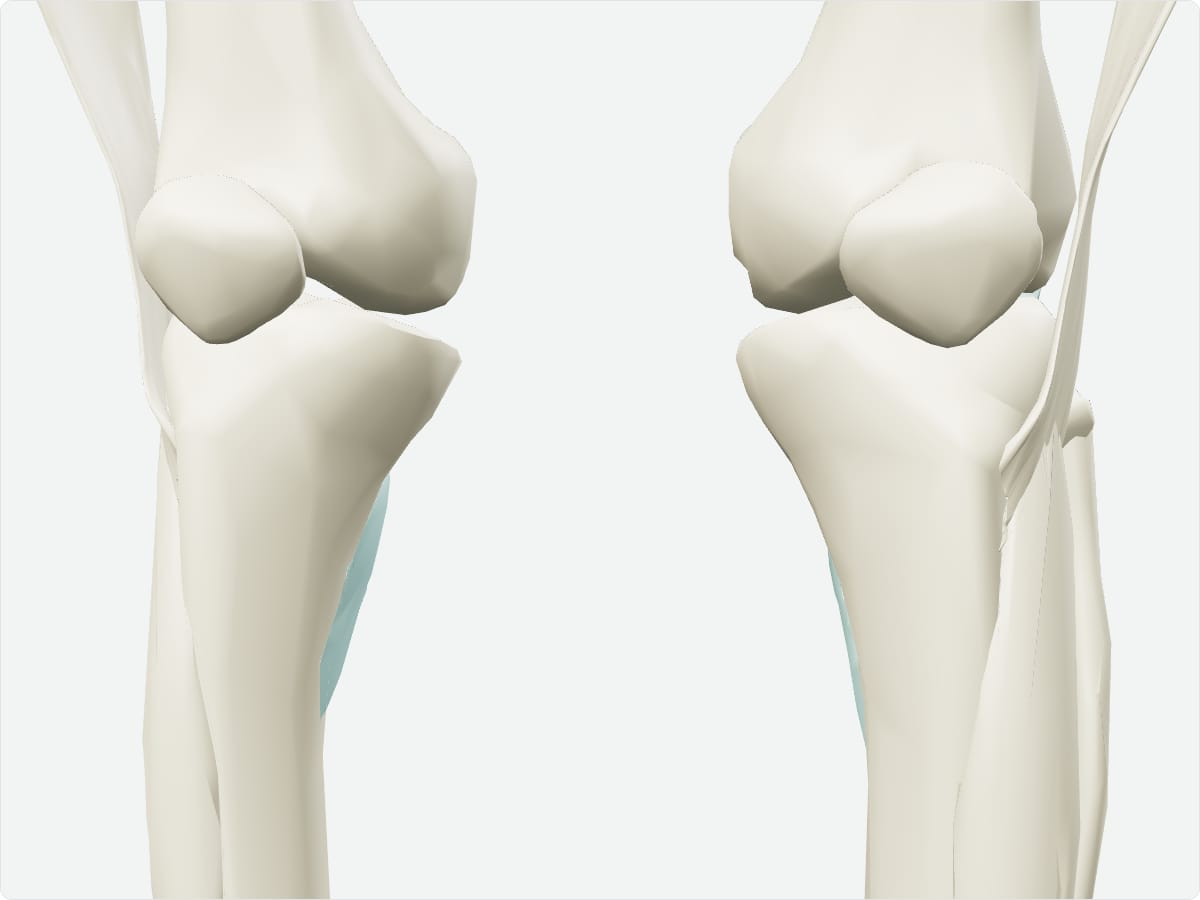
3D model: BodyParts3D (CC BY 4.0), adult male reference anatomy. Highlighted structure shown with nearby bones for context.
About the popliteus
The popliteus forms part of the floor of the popliteal fossa. Its tendon passes inside the knee joint capsule, beneath the lateral collateral ligament.
Clinical relevance
Popliteus injury is often associated with posterolateral corner injuries of the knee.
Frequently asked questions
What does the popliteus do?
It unlocks the extended knee so that flexion can begin.