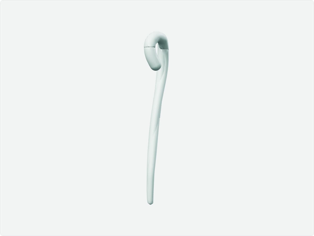
The aorta is the largest artery of the body. It arises from the left ventricle and includes the ascending aorta, aortic arch, descending thoracic aorta and abdominal aorta, ending at L4 where it divides into the common iliac arteries. The arch gives off the brachiocephalic trunk, left common carotid and left subclavian arteries.
Key facts
Location
From the left ventricle through the mediastinum, then through the diaphragm at T12 into the abdomen.
Structure
Ascending aorta, aortic arch, descending thoracic aorta and abdominal aorta; aortic root with sinuses.
Function
Distributes oxygenated blood from the heart to the systemic circulation.
Blood supply
Vasa vasorum supply the outer wall; the coronary arteries arise from the aortic root.
Innervation
Aortic plexus; baroreceptors in the arch relay to the vagus nerve.
3D model: BodyParts3D (CC BY 4.0), adult male reference anatomy. Highlighted structure shown with nearby bones for context.
About the aorta
Branches of the arch supply the head, neck and arms. The abdominal aorta supplies the digestive organs, kidneys and pelvis via celiac, mesenteric and renal branches.
Clinical relevance
Abdominal aortic aneurysm is a dilation of the vessel that can rupture without warning.
Aortic dissection is a tear in the inner layer that requires emergency treatment.
Frequently asked questions
What are the branches of the aortic arch?
The brachiocephalic trunk, left common carotid artery and left subclavian artery.
Where does the aorta divide?
At about L4, into the right and left common iliac arteries.